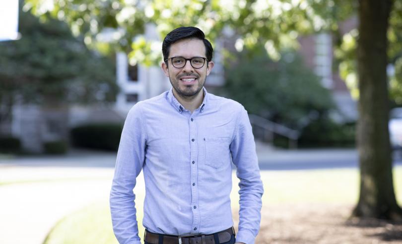
Physics & Astronomy Colloquium

Dr. Stephen Taylor, Vanderbilt University

Title: Charting the Gravitational-wave Universe At Light-year Wavelengths

Abstract: The Universe is thrumming with gravitational waves. June 2023 brought the first evidence for an all-sky background of nanohertz-frequency gravitational waves, discovered by collaborations including the North American Nanohertz Observatory for Gravitational Waves (NANOGrav) and groups in Europe, Australia, India, and China. This was an endeavor decades in the making, requiring painstakingly precise timing observations of scores of millisecond pulsars across the Milky Way using flagship radio telescopes. While the results from separate groups are consistent with one another—and the leading interpretation of a population of supermassive black-hole binaries as the source—the observations provoke many new questions. Do the results imply a population of binaries more massive than expected? What are the observational milestones as the first individually resolvable binary signals come into focus? Can we link these signals to their host galaxies or electromagnetic counterparts? In this talk, I will chart the path to discovery, reflect on what we have learned since our announcement, and explore the exciting opportunities and synergies ahead—including the role of next-generation radio instruments and space-borne gravitational-wave missions.

Date:
-
Location:
CP 153
Event Series:

Physics & Astronomy Colloquium

Dr. Zhoudunming (Kong) Tu, Brookhaven National Laboratory

Title: Exciting the Entangled Vacuum - A New Era in Understanding Visible Matter

Abstract: Not until quite recently was the vacuum recognized as anything more than empty space. Today, we understand it as a dynamic medium, filled with fluctuating fields and virtual particle pairs that shape the very structure of our universe. These invisible pairs break a fundamental symmetry of nature—chiral symmetry—and are thought to generate more than 99% of the mass of the visible universe. Yet, how this hidden mechanism connects to the confinement of quarks inside protons, neutrons, and other particles remains one of the deepest unsolved problems in physics.

In this talk, I will present new insights into this question using high-energy particle collisions at the Relativistic Heavy Ion Collider (RHIC). Such collisions can briefly liberate the virtual quark–antiquark pairs of the vacuum, which then bind together into hadrons such as Λ hyperons. Recent results from these studies open an experimental window into the quantum structure of the vacuum, with far-reaching implications for our understanding of mass, matter, and the strong force described by Quantum Chromodynamics.

Date:
-
Location:
CP 153
Event Series:

"Space Medicine and the Future of Human Exploration"

A self photo of Dr. Afshin Beheshti in a navy blue collared button up shirt.
Dr. Afshin Beheshti 
 

Bio:
Afshin Beheshti, PhD is a Professor of Surgery and of Computational & Systems Biology at the University of Pittsburgh School. He serves as Director of the newly launched Space Center for Space Biomedicine and as Associate Director of the McGowan Institute for Regenerative Medicine at Pitt​. In addition, Dr. Beheshti holds a visiting scientist appointment at the Broad Institute of MIT and Harvard​.

Abstract:
Human spaceflight presents significant health challenges driven by microgravity, space radiation, isolation, and other environmental stressors. Recent multi-omics research has revealed that mitochondrial dysfunction is a central biological consequence of space travel, contributing to systemic impacts such as accelerated aging, cardiovascular disease, and impaired metabolic function. Data from astronaut missions and ground-based space analogs demonstrate persistent mitochondrial suppression even after returning to Earth. This talk highlights how space serves as a unique accelerated model for studying human diseases and aging, offering insights applicable both to space exploration and terrestrial medicine. Using advanced 3D organoid models and multi-omics analysis, we have identified promising countermeasures, including the natural flavonoid Kaempferol, which restores mitochondrial bioenergetics and reverses radiation-induced gene expression changes in multiple tissues. These findings underscore the critical role of mitochondria as both biomarkers and therapeutic targets for sustaining human health in deep space missions, while also advancing precision medicine strategies on Earth.

Date:
Location:
THM 116

"Understanding the Sources of Regenerative Capacity in Animals"

Dr. Alejandro Sánchez Alvarado standing in front of a white dry erase board with his arms crossed while smiling. The dry erase board behind him has biology graphics in different colors.

Alejandro Sánchez Alvarado | Sánchez Alvarado Lab

Bio:
Sánchez Alvarado received a BS in molecular biology and chemistry from Vanderbilt University in Nashville, Tennessee, and a Ph.D. in pharmacology and cell biophysics from the University of Cincinnati College of Medicine in Cincinnati. He performed postdoctoral and independent research at the Carnegie Institution of Washington, Department of Embryology in Baltimore. In 2002, he joined the faculty of the University of Utah School of Medicine in Salt Lake City, where he held the H.A. & Edna Benning Presidential Endowed Chair. In 2005, he was named a Howard Hughes Medical Institute investigator. He joined the Stowers Institute for Medical Research in Kansas City in 2011 and became the president and chief scientific officer of the Stowers Institute in 2022. He also holds the Priscilla Wood Neaves Chair in the Biomedical Sciences.

Sánchez Alvarado is an elected member of the National Academy of Science, the American Academy of Arts and Sciences and the Latin American Academy of Sciences; a Kavli Fellow of the National Academy of Sciences USA; a fellow of the Marine Biological Laboratory in Woods Hole, MA; a fellow of the American Association for the Advancement of Science; and a recipient of a National Institutes of Health MERIT award, the EE Just Medal for Scientific Achievement and the Vilcek Prize in Biomedical Sciences. He has served on numerous scientific advisory committees and boards including the National Advisory Council of the National Institute of General Medical Sciences and the National Institutes of Health. He serves on the Board of Directors of American Century Investments.

Sánchez Alvarado’s work has the potential to lead to a better understanding of how the adult forms of higher organisms, including humans, carry out their biological functions. His research also has led to insights on the molecular and genetic drivers of both regenerative and degenerative cellular processes that contribute to disease.

Abstract:
It is paradoxical that for many organisms (including humans), the apparent anatomical stability of their adult bodies is maintained by constant change. Despite the importance of tissue homeostasis and regeneration to human biology and health, relatively little is known about how these processes are regulated. As such, numerous questions remain unanswered, including: How do organ systems maintain their order and function while in a state of cell flux? How do animals control and coordinate the size and cell number of multiple organ systems? Does regeneration of body parts lost to injury invoke embryonic processes, generic patterning mechanisms, or unique circuitry comprised of well-established patterning genes? Answering any of these questions would set a baseline from which to try to enhance regenerative properties in multicellular organisms such as humans, particularly after injury.

One way to solve a complex problem is to reduce it to a simpler, easier to answer problem. Therefore, reducing the complexities of regeneration and tissue homeostasis to the study of comparatively simpler systems would allow for a systematic dissection and mechanistic understanding of these processes. Here, I will discuss how the use of single-cell and spatial transcriptomics is helping define the cellular and molecular environments that support pluripotency in the highly regenerative freshwater planarian Schmidtea mediterranea and regeneration of missing organs in the hemichordate Ptychodera flava. Our studies are beginning to shed light on the way adult animals regulate tissue homeostasis and the replacement of body parts lost to injury.

Date:
Location:
THM 116

"Orchestrating Mammalian Tissue Healing at the Organ Scale"

Image of Dr. Yvon WoappiDr. Yvon Woappi | Woappi Lab

Bio:
Dr. Yvon Woappi is the Herbert and Florence Irving Assistant Professor of Physiology and Cellular Biophysics, Dermatology, and Biomedical Engineering at Columbia University. His research leverages gene editing and multiomic technologies to uncover how autonomous multicellular orchestration facilitates deep wound repair – a process critical to many conditions including diabetic ulcers and carcinomas. Dr. Woappi earned his Ph.D. as a Grace Jordan McFadden Fellow at the University of South Carolina and completed his postdoctoral training in the Harvard Dermatology Research Training Program at Harvard Medical School. Dr. Woappi’s pioneering early-career research is laying the foundation for synthetic wound regeneration, a systems bioengineering approach that leverages cellular heterogeneity to enhance tissue regeneration.

Abstract:
As the organ most frequently exposed to predatory pressures, the integument has acquired broad functions, including camouflage, thermoregulation, sensory perception, and tissue repair. These roles are executed through a complex interplay of tissue substructures, including several mini-organ appendages (hair follicles, sebaceous glands, arrector pili muscle, and assorted pilosebaceous units) and five central adnexal structures (blood vessels, sensory neurons, collagenous tissues, immune components, and deep fascia), all embedded within three superimposed tissue strata (the epidermis, dermis, and hypodermis). Given this intricate architecture, the healing of deep skin wounds requires a coordinated organ-level response involving varied cell populations originating from virtually all three embryonic germ layers—ectoderm, mesoderm, and endoderm. However, a comprehensive understanding of the cellular and molecular logic orchestrating this crosstissue response in mammals remains incomplete. Here, we present the Organ-Scale Wound Healing Atlases (OWHA), a comprehensive multiomic single-cell and spatial transcriptomic dataset that captures the dynamic microanatomical tissue niches of the mammalian integument during the entire wound healing sequence, including early and late healing phases. By incorporating multi-omics data across all major phases of healing, OWHA uncovered novel emergent healing cell states and their coordinated cell fate decisions uniquely (multilineage crosstalk) executed after injury, and delineated critical tissue trajectories required for eKective healing of deep wounds. Importantly, comparative analysis between human and mouse revealed conserved network between the epithelial and neuro-endothelial vasculature....(Add missing groups in human only found in our multi modal approach) By providing deeper mechanistic insights of mammalian tissue adaptations for injury response, OWHA serves as a valuable resource for understanding the cellular and molecular mechanisms underlying wound healing in the mammalian integument.

A picture of mouse skin with hair follicles.

Date:
Location:
THM 116

"Of Microbes and Microglia: Sex-Biased Immune Pathways in Stress and Mood Disorders"

Dr. Georgia Hodes standing in front of a blue tie-dyed like background smiling. She's wearing a black dress with pink and red flowers throughout. She's also wearing an off gold necklace with red flowers.Dr. Georgia Hodes 

Bio:
Dr. Hodes received a B.A. in Drama/Dance from Bard College and after college worked as an actor and designer in New York City. During this time, she decided a life in the arts was unsustainable and did post-baccalaureate training at Hunter College in experimental psychology. She obtained her Ph.D. from Rutgers University in the Behavioral and Systems Neuroscience division of the Psychology program where she trained in the laboratory of Dr. Tracey Shors. She went on to have 2 postdoctoral training positions, the first in Pharmacology with Dr. Irwin Lucki at the University of Pennsylvania and the second in Neuroscience with the Dr. Scott Russo at the Icahn School of Medicine at Mt. Sinai. She has received 2 NARSAD young investigator awards and is an author on over 70 publications. In 2016 she joined the faculty of the newly formed School of Neuroscience at Virginia Tech. Her research program examines sex differences in the peripheral and central immune system and how immune mechanisms interact with brain plasticity to drive behavioral differences in emotional processing of stress and mental health disorders.

Abstract:
The brain does not exist in a vacuum. The brain controls the body; however, the body also impacts the brain. This occurs through a variety of immune mechanisms, including cytokines and microbes produced in the periphery. When we get sick, our cells produce or suppress signals depending on the type of invasion. They tailor the environment to kill off an invading pathogen or our own injured cells. Cells then produce other signals to shut down this response and promote recovery and healing.  Like physical illness, mental illness, or even stress exposure, alters immune signaling in both the body and the brain. The fundamental question driving my research is how immune pathways contribute to the pathology associated with the development of mental illness, and why are some individuals more vulnerable than others to this type of immune dysfunction? 

Watch the seminar here!

Date:
Location:
THM 116

"An Active Role for Vision Prior to Eye-opening in Neonates"

Photo of Dr. D'Souza smiling in a white lab coat. In the background you can see blurred green leaves from a tree in the back right corner and the side of a building in the back left corner.Dr. Shane D'Souza

Bio:
Dr. Shane Peter D’Souza is a neuroscientist and vision researcher whose work spans developmental neurobiology, sensory physiology, and circadian biology. He earned his BS in Biology at the University of Kentucky and PhD in Molecular and Developmental Biology from the University of Cincinnati/Cincinnati Children’s Hospital Medical Center, where he investigated how early light exposure shapes neural circuit development in the retina and brain. Now a Postdoctoral Research Fellow in Pediatric Ophthalmology at Cincinnati Children’s, Dr. D’Souza’s research integrates molecular, anatomical, physiological, and computational approaches to understand how intrinsically photosensitive retinal ganglion cells (ipRGCs) and other non-visual photoreceptors influence sensory-driven circuit refinement, photoreceptor abundance, and cross-modal communication in early life. His publication record includes discoveries on melanopsin-dependent regulation of rod photoreceptors, neuropsin and encephalopsin function in visual and non-visual systems, and the role of light-sensitive hypothalamic neurons in thermoregulation and metabolism. By combining high-resolution imaging, transcriptomics, biophysical modeling, and machine-learning, his work aims to uncover fundamental principles of sensory-mediated neural development and sensory adaptation across species. In his spare time, he enjoys studying the evolution of storm systems and serves as a storm spotter for NWS Wilmington, OH. In his spare-spare time, he writes and produces music from his living room.

Abstract:
Most mammals are born with immature, poorly developed sensory systems. As these systems come online, they use immature sensory experience to shape synapses, cell types, and their connectivity across the brain. In the visual system, this experience is thought to be passive, supporting and setting up later modes of image-forming vision after eye-opening.  However, little is known about the form and function of visual experience during the earliest period in a neonate’s life. Driven by this, we set out to generate a comprehensive map of visual system activity and neonatal behaviors in mice.  Using a host of machine learning-based approaches we developed an atlas perinatal visual system activity from the retina to several regions of the brain. Using a combination of chromatic stimuli and genetic loss-of-function mice, we identified the M1 intrinsically photosensitive retinal ganglion cell (ipRGC) in the retina as the driver of early visual system activity, activating distinct brain regions during development. Using this atlas as a guide to assess behaviors, we find that this visual input drives the production of ultrasonic vocalization in neonates and “blinding” mice leads to an augmented vocal code.  Together, these data suggest that early visual system activity has an active role in supporting the development of neonatal behaviors and warrants a deeper exploration of early sensory activity across the developing brain.

Watch the seminar here!

A photo of Dr. D'Souza in a white lab coat crouched in some grass holding a doggie toy. Across from Dr. D'Souza is a Golden Retriever with a harness on and it's tongue hanging out of its mouth.Pictured is a color enhanced mouse brain.


Date:
Location:
THM 116

"Old and New: Trait Evolution in Natural Populations"

Dr. Molly Schumer leaned up against a stone wall with one arm across her waist holding the other arm while smiling.Dr. Molly Schumer | Schumer Lab

Bio:
Molly Schumer is an Assistant Professor in Biology. She is interested in genetics and evolutionary biology. After receiving her PhD at Princeton, she did her postdoctoral work at Columbia and was a Junior Fellow in the Harvard Society of Fellows and Hanna H. Gray Fellow at Harvard Medical School. Current research in the lab centers on understanding the genetic mechanisms of evolution, with a focus on natural populations.

Abstract:
The diverse branches of life on earth trace to a common root. In the past two decades, a revolution in genome sequencing has allowed researchers to make unprecedented progress in understanding the evolution of life on earth at the genetic level. Our lab is interested in why and how new species arise, and what genetic changes underlie their ability to adapt to the environments in which they live. To study these questions, we use an interdisciplinary approach – melding genomics and evolution with molecular biology, behavior, and physiology. Our work leverages an emerging model system, swordtail fish or Xiphophorus, where we can study genetics and evolution  using experimental and natural populations.

Date:
Location:
THM 116

"Adaptation in Arabidopsis through Space and Time"

Pictured is Dr. Jesse Lasky wearing a plaid collared navy with pink and yellow button up shirt.Dr. Jesse Lasky | Lasky Lab

Bio:
Dr. Jesse Lasky attended Kenyon College and received his BA in Biology and a PhD in Ecology Evolution & Behavior at UT-Austin. Dr. Lasky was a postdoc at Columbia University and is now a faculty member at Penn State University, in addition to being the herbarium director.

Abstract:
Global patterns of population genetic variation through time offer a window into evolutionary processes that maintain diversity. Over time, lineages may expand or contract their distribution, causing turnover in population genetic composition. At individual loci, migration, drift, and selection (among other processes) may affect allele frequencies. Museum specimens of widely distributed species offer a unique window into the genetics of understudied populations and changes over time. Here, we sequenced genomes of 130 herbarium specimens and 91 new field collections of Arabidopsis thaliana and combined these with published genomes. We sought a broader view of genomic diversity across the species, and to test if population genomic composition is changing through time. We documented extensive and previously uncharacterized diversity in a range of populations in Africa, populations that are under threat from anthropogenic climate change. Through time, we did not find dramatic changes in genomic composition of populations. Instead, we found a pattern of genetic change every 100 years of the same magnitude seen when comparing Eurasian populations that are 185 km apart, potentially due to a combination of drift and changing selection. We found only mixed signals of polygenic adaptation at phenology and physiology QTL. We did find that genes conserved across eudicots show altered levels of directional allele frequency change, potentially due to variable purifying and background selection. Our study highlights how museum specimens can reveal new dimensions of population diversity and show how wild populations are evolving in recent history.

Watch the seminar here!

A photo with dried Arabidopsis thaliana, also known as thale cress, taped to an off white sheet of paper. There is a bright white square piece of paper in the bottom left corner that has writing. The title of this sheet of paper is "Herbier du Bureau de Botanique appliquee, Petrograd.".

Date:
Location:
THM 116
Subscribe to